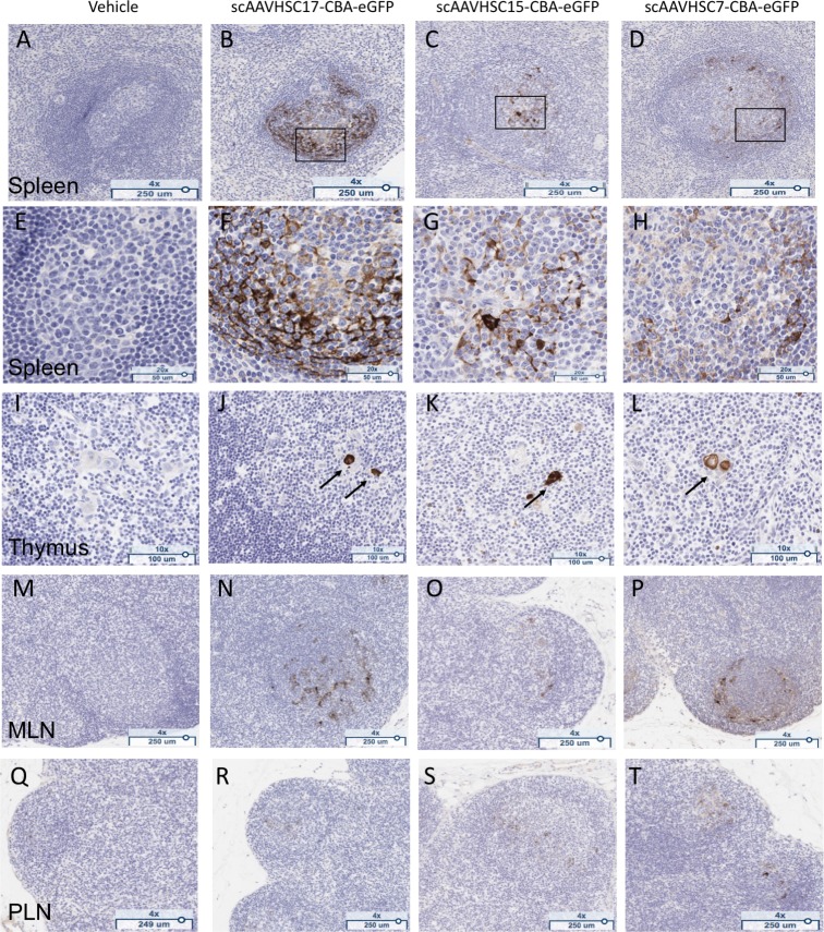
Fig 18. eGFP detection in lymphoid tissue of nonhuman primates treated with scAAVHSC-CBA-eGFP.
(A, E, I, M, Q) The animal received an IV dose of vehicle alone. (B, F, J, N, R) The animal received an IV dose of scAAVHSC17-CBA-eGFP. (C, G, K, O, S) The animal received an IV dose of scAAVHSC15-CBA-eGFP. (D, H, L, P, T) The animal received an IV dose of scAAVHSC7-CBA-eGFP. Tissues were isolated two weeks post-dose for eGFP staining as described under Materials and methods. White pulp splenic nodules are shown in A-D with higher magnification views of the boxed areas shown in F-H. eGFP staining in Hassall’s bodies (arrows) of the thymic medulla are shown in I-L, and eGFP staining within the germinal centers of mesenteric (MLN) and peripheral (PLN) lymph nodes are shown in M-P and Q-T, respectively. Brown staining represents eGFP staining. Representative tissues are shown.

