. 2019 Nov 5;8:e48940. doi: 10.7554/eLife.48940

Figure 2. AAGAG RNA is enriched in primary spermatocytes and necessary for male fertility.

(a) Confocal section of a larval testis. RNA-FISH to AAGAG = magenta, H2Av (chromatin) IF = gray, DNA (DAPI) = blue. S3, S5, and S6 refer to primary spermatocyte stages. (b) Enlarged confocal sections (representative boxes in a) of spermatocyte stages in larvae testes; scale bars = 5 µm. (c) Schematic summary of AAGAG RNA (magenta) localization in adult testes (see Figure 2—figure supplement 1 for a detailed description of spermatogenesis stages and events). AAGAG RNAs are visible in 16 cell primary spermatocytes (dark pink), and potentially 16 cell spermatogonial cysts (light pink); no AAGAG RNA was detected at earlier stages (hub, 2–8 cell spermatogonial cysts) or after the primary spermatocyte stage (meiosis I and II, sperm elongation- which includes leaf, canoe, individualization steps, and maturation). Post-round spermatid stages are indicated as spermatid nuclei. (d) Fertility after depletion of AAGAG(n) RNA in male primary spermatocytes or female ovaries using the Bam-GAL4 driver. An ~72% reduction in AAGAG RNA levels in testes (see Figure 2—figure supplement 3, B and C) results in complete male sterility but has no effect on female fertility. Expression of AAGAG(37) RNA simultaneously with AAGAG RNAi (both driven by Bam-Gal4) partially rescues male sterility (46% fertile). Expression of AAGAG RNA alone, without depletion of endogenous AAGAG RNAs, has no impact on male fertility. Statistically significant differences based on T-tests (two tailed, type three) are indicated by horizontal lines; ***p<0.001, **p<0.01; variation is represented by stdev.

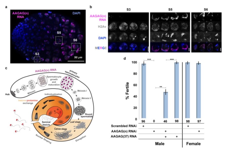
Figure 2.

Figure 2—figure supplement 1. Overview of normal spermatogenesis and defects observed after AAGAG RNA depletion.

Figure 2—figure supplement 1.

(a) Spermatogenesis in Drosophila melanogaster initiates at the apical end of the testes (Hub), where GSCs divide asymmetrically, producing gonialblasts (GBs) that begin cell-differentiation. GB cells then undergo four mitotic divisions with incomplete cytokinesis to produce a cyst of 16 primary spermatocytes. Spermatocytes then undergo pre-meiotic S phase, mature during a prolonged G2 phase, and increase substantially in volume. The majority of testes-specific gene expression occurs at the primary spermatocyte stage, while genes not required until later stages are translationally repressed (reviewed in White-Cooper, 2010). Mature spermatocytes then undergo two rounds of meiosis to produce round spermatids (McKee et al., 2012), which are then processed into independent, condensed sperm nuclei in two stages (Rathke et al., 2014; Eren-Ghiani et al., 2015; Steinhauer, 2015). First, round spermatids undergo chromatin compaction, acrosome formation and flagellar elongation (Rathke et al., 2014; Eren-Ghiani et al., 2015). During chromatin compaction, a wave of histone H4 acetylation occurs, followed by deposition of the transition protein Mst77f, (Kost et al., 2015). Next, transition proteins are removed followed by the incorporation of protamines and prtl99c (histone:protamine exchange, indicated by tan to deep orange gradient) (Rathke et al., 2014; Eren-Ghiani et al., 2015). Finally, spermatid individualization involves removal of cytoplasm and tight condensing and coiling of chromatin (Steinhauer, 2015). Mature sperm are then stored in the seminal vesicle. (b) Summary of defects in late stages of spermatogenesis observed after depletion of AAGAG RNA by RNAi, using the Bam-Gal4 driver (data in Figure 3). Although AAGAG RNA is not visible in normal testes after the S6 spermatocyte stage (see a), RNAi depletion of AAGAG RNA only produces visible defects after the round spermatid stage. Aberrant elongation, sperm bundles, and defective histone:protamine exchange likely cause the observed complete absence of mature sperm in the SV.

Figure 2—figure supplement 2. Heterochromatic regions adjacent to AAGAG(n) or AG(n)-rich blocks are transcribed in primary spermatocytes, co-localize with AAGAG(n) RNA foci and do not come from the Y.

Figure 2—figure supplement 2.

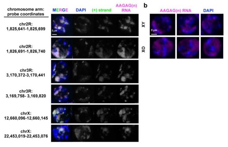
(a) Projections of Oregon R S5 spermatocytes probed for unique regions of RNA (green) adjacent to AAGAG(n) (magenta) or AAGAG(n) containing AG rich blocks. DAPI (DNA) is indicated in blue. (b) Projections of S5 spermatocyte probed to AAGAG RNA (magenta) imaged at same laser intensities in XY and XO genotypes. DNA is stained with DAPI (blue).

Figure 2—figure supplement 3. AAGAG RNA and not CUCUU RNA is substantially decreased in Bam-GAL4- driven AAGAG RNAi, and AAGAG RNA levels are increased in rescue experiments.

Figure 2—figure supplement 3.

(a) Although visibly absent in embryos and somatic larval tissues, CUCUU RNA (green) is expressed in adult spermatocytes. Note that CUCUU RNA is localized to the S5 lumen, internal to the chromatin (DAPI), in contrast to the peripheral localization of AAGAG RNA (see Figure 3b); DNA = DAPI (blue). (b) Projections of AAGAG foci (magenta) in S5 spermatocytes after Bam-GAL4-driven Scrambled control or AAGAG RNAi. Signal was imaged with the same laser intensities for each genotype. (c) Average median intensities (arbitrary units, ± st. dev.) of AAGAG RNA, p=2×10⁻⁵ and CUCUU RNA in S5 spermatocytes in AAGAG and Scrambled RNAi testes (not significant). This represents a 72% reduction of AAGAG RNA in S5 spermatocytes after AAGAG RNAi, compared to scrambled controls, with little to no decrease in CUCUU RNA. (d) Average intensity of AAGAG RNA in S5 spermatocytes after AAGAG RNAi increases significantly (p=0.03) upon co-expression of AAGAG(37) RNA (also induced by the Bam-Gal4 driver). two tailed, type three t test used for all.