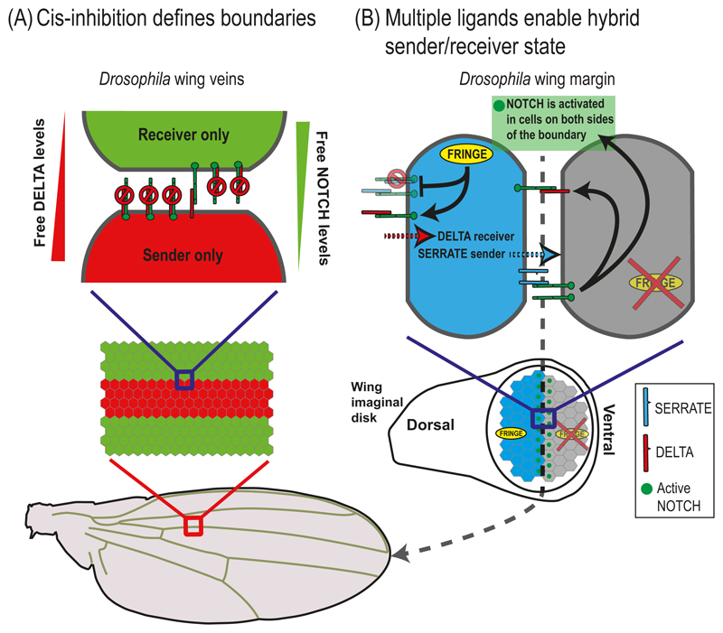
Fig. 5.4. NOTCH signaling in boundary formation.
(A) Cis-inhibition defines boundaries. A schematic overview of the cis-inhibition model for the wing vein boundary in Drosophila. In this model, wing vein boundaries are defined by the interactions between "sender" cells and "receiver" cells. The cells in the vein region (red) express more DELTA than NOTCH while the cells in the inter-vein region (green) express more NOTCH than DELTA. Due to cis-interactions between NOTCH receptors and ligands, vein cells can send but not receive signals, while inter-vein cells can receive but not send signals. (B) Multiple ligands enable hybrid sender/receiver states. A schematic model for wing margin cells in Drosophila taking into account multiple ligands and modulation by FRINGE. The cells on the dorsal side (blue) express both DELTA, SERRATE and FRINGE. The FRINGE glycosyltransferase modulates the cis- and trans-interactions between NOTCH receptors and ligands. Expression of FRINGE promotes NOTCH-DELTA interactions and suppresses NOTCH-SERRATE interactions (both in cis and in trans). The model predicts that the dorsal boundary cells (blue) can simultaneously receive signals from DELTA expressing cells and send signals to the ventral boundary cells (gray) using the SERRATE ligands. At a later stage, the ventral boundary cells activate DELTA leading to NOTCH activation in the dorsal boundary cells. This situation leads to activation of cells only on the wing margin (green dots in bottom image).

