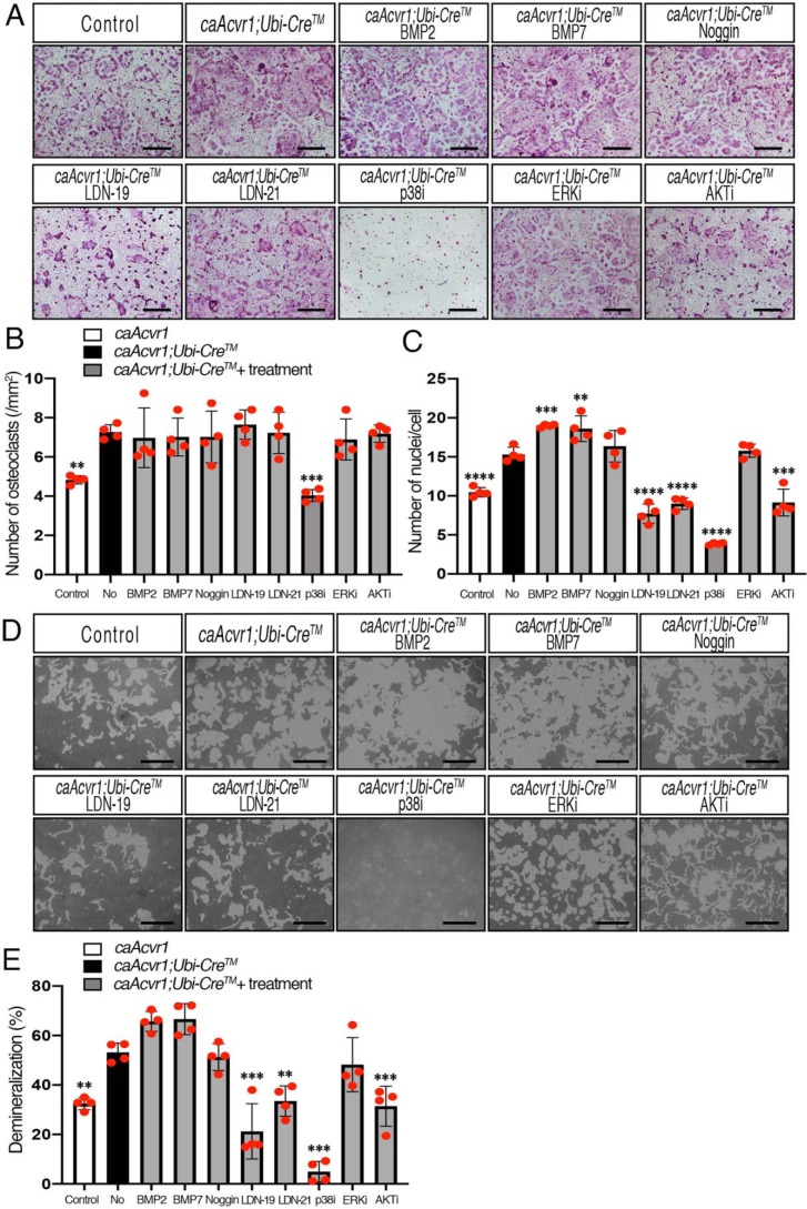
Figure 6.
LDNs inhibited osteoclast fusion and activity of caAcvr1-mutant cells. A, BMMs from control (caAcvr1) and caAcvr1-mutant (caAcvr1;Ubi-CreTM) mice were seeded in 48-well plates (1.5 × 10⁴ cells/well) with M-CSF (20 ng/ml), RANKL (100 ng/ml), and 4-hydroxytamoxifen (100 ng/ml) for 5 days and TRAP staining was conducted. caAcvr1-mutant cells were treated with DMSO (0.1%, as nontreatment), BMP-2 (100 ng/ml), BMP-7 (100 ng/ml), Noggin (250 ng/ml), LDN-19 (LDN-193189, 1 μm), LDN-21 (LDN-212854, 1 μm), p38 MAPK inhibitor (SB203580, 1 μm), ERK inhibitor (UO126, 1 μm), and PI3K/AKT inhibitor (LY294002, 1 μm). Control cells were treated with DMSO at a final concentration of 0.1%. The value of each group was compared with the nontreatment group. Scale bars, 500 μm. B, TRAP-positive cells containing three or more nuclei were counted as osteoclasts. n = 4. C, number of nuclei per cell was analyzed. n = 4. D, cells were incubated with M-CSF (20 ng/ml), RANKL (100 ng/ml), and 4-hydroxytamoxifen (100 ng/ml) for 7 days. The demineralized pits generated by osteoclasts were visualized by von Kossa staining. Scale bars, 500 μm. E, demineralized area was measured using ImageJ. The value of each group was compared with nontreatment group of caAcvr1-mutant cells. n = 4. Values represent the mean ± S.D. Differences were assessed by one-way ANOVA, followed by a Dunnett test, **, p < 0.01; ***, p < 0.001; ****, p < 0.0001.

