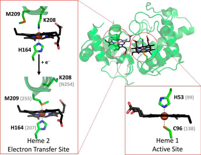
Figure 2.

The structure of A. vinosum TsdA with the two heme cofactors highlighted in black. Expanded regions show the ligands to Heme 1 and those to Heme 2 in the oxidized (Fe(III)) and reduced (Fe(II)) states. Carbon atoms are shown in green, nitrogen in blue, oxygen in red and sulfur in orange. Where appropriate, gray values in parentheses indicate the corresponding residues from C. jejuni TsdA, as deduced from sequence alignment. Reproduced from PDB entries 4WQ7 and 4WQ9.
