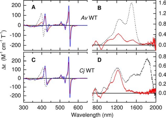
Figure 4.

A–D, MCD spectra of A. vinosum (A and B) and C. jejuni (C and D) TsdA following incubation with sodium ascorbate (red lines) and sodium dithionite (blue lines). Protein concentrations were 35 and 20 μm (ascorbate and dithionite respectively, 300–700 nm) and 178 μm (700–2000 nm) for AvTsdA and 37 and 36 μm (300–700 nm) and 145 μm (700–2000 nm) for CjTsdA. The broken lines are spectra of the fully oxidized enzymes from Fig. 3 for comparison. Data recorded at room temperature in 50 mm HEPES, 50 mm NaCl, pH 7, or the same buffer in D2O pH* 7 for nIR MCD.
