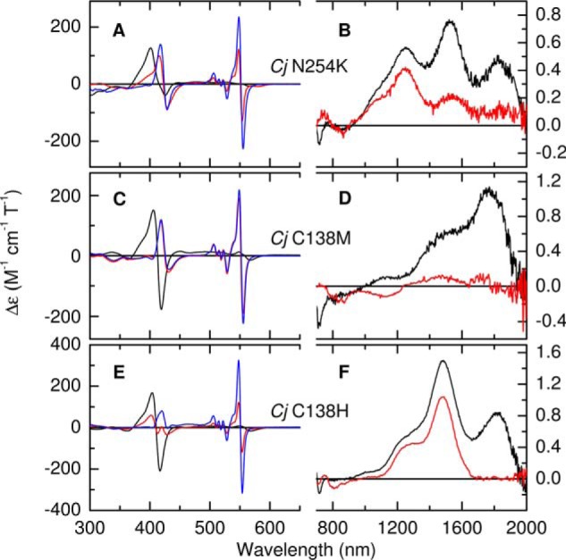
Figure 7.

A–F, the MCD spectra of CjTsdA variants N254K (A and B), C138M (C and D), and C138H (E and F), in the fully oxidized state (black traces), following incubation with sodium ascorbate (red traces) and with sodium dithionite (blue traces). Protein concentrations used for the 300–650 nm and the 700–2000 nm regions were, respectively, 47 μm and 325 μm; 40 μm and 160 μm; 18 μm and 169 μm. Data recorded at room temperature in 50 mm HEPES, 50 mm NaCl, pH 7, or the same buffer in D2O pH* 7 for nIR MCD.
