Figure 1.
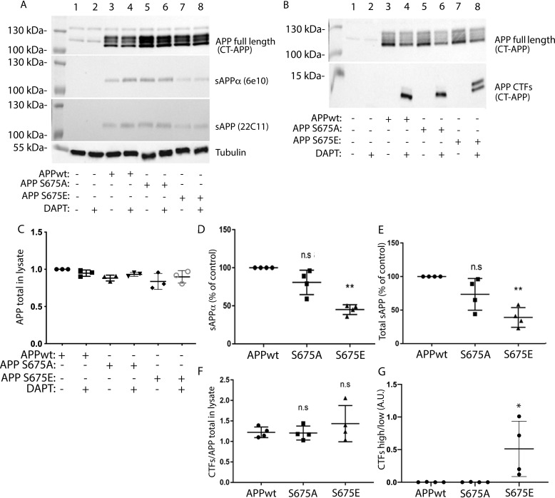
APP–Ser-675 phosphorylation decreases sAPPα secretion while increasing the level of a slower migrating APP-CTF. A, representative Western blot analysis of full-length APP (detected by CT-APP), sAPPα (detected by 6E10), and total sAPP (detected by 22C11) from nontransfected (lanes 1 + 2) or APPwt (lanes 3 + 4), APP-S675A (lanes 5 + 6), or APP-S675E (lanes 7 + 8) transfected SK-N-AS cells, in the absence (lanes 1 + 3 + 5 + 7) or presence (lanes 2 + 4 + 6 + 8) of the γ-secretase inhibitor DAPT. B, representative Western blot analysis of APP-CTFs (detected with CT-APP) from nontransfected (lanes 1 + 2) or APPwt (lanes 3 + 4), APP-S675A (lanes 5 + 6), or APP-S675E (lanes 7 + 8) transfected SK-N-AS cells, in the absence (lanes 1 + 3 + 5 + 7) or presence (lanes 2 + 4 + 6 + 8) of the γ-secretase inhibitor DAPT. C, quantification of the full-length APP level, normalized against the corresponding tubulin level. D and E, relative abundance of secreted sAPPα and total sAPP in culture medium from DAPT-treated APPwt, APP-S675A, and APP-S675E overexpressing SK-N-AS cells. The level of sAPPα and total sAPP were normalized against both the level of corresponding total APP expression and the protein concentration in cell lysate. F, quantification of the total APP-CTF levels, normalized against the corresponding APP full-length level, in cell lysate from DAPT-treated APPwt, APP-S675A, and APP-S675E overexpressing SK-N-AS cells. G, ratio of APP-CTF upper/APP-CTF lower band in cell lysate of DAPT-treated APPwt, APP-S675A, and APP-S675E overexpressing SK-N-AS cells. For quantifications, *, p < 0.05; **, p < 0.01; n.s, not significant; n = 3–4.

