Figure 6.
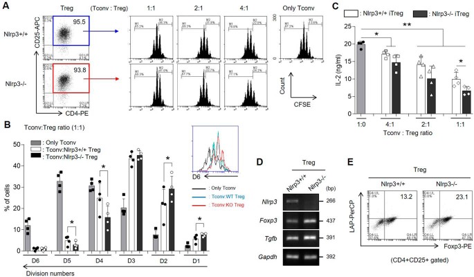
Nlrp3−/− iTreg populations are functionally more suppressive than Nlrp3+/+ iTregs. A–C, the iTreg-mediated suppression activity was measured by CFSE dilution of Tconv cells. The CFSE-stained Tconv cells were co-cultured for 3 days with Nlrp3+/+ or Nlrp3−/− iTregs sorted as CD4+CD25+ T cells by at various ratios, or cultured alone. A, proliferation of Tconv cells was determined by CFSE dilution and flow cytometric analysis. B, the percentages of divided Tconv cells were arranged by splitting the number of division in the presence of either Nlrp3+/+ iTregs or Nlrp3−/− iTregs at a 1:1 of Tconv: iTreg ratio. C, the secretory level of IL-2 from Tconv cells was measured by ELISA. D, the mRNA expression levels of Nlrp3, Tgfb as well as Foxp3 were detected in both WT and KO iTregs after Treg differentiation for 72 h. E, the population of Foxp3+LAP (latent TGF-β)+ cells in CD4+CD25+ iTregs were assessed by flow cytometry. Data are representative of four independent experiments (n = 4). The results are presented as mean ± S.D. *, p < 0.05; **, p < 0.01 (Student's two-tailed t test).

