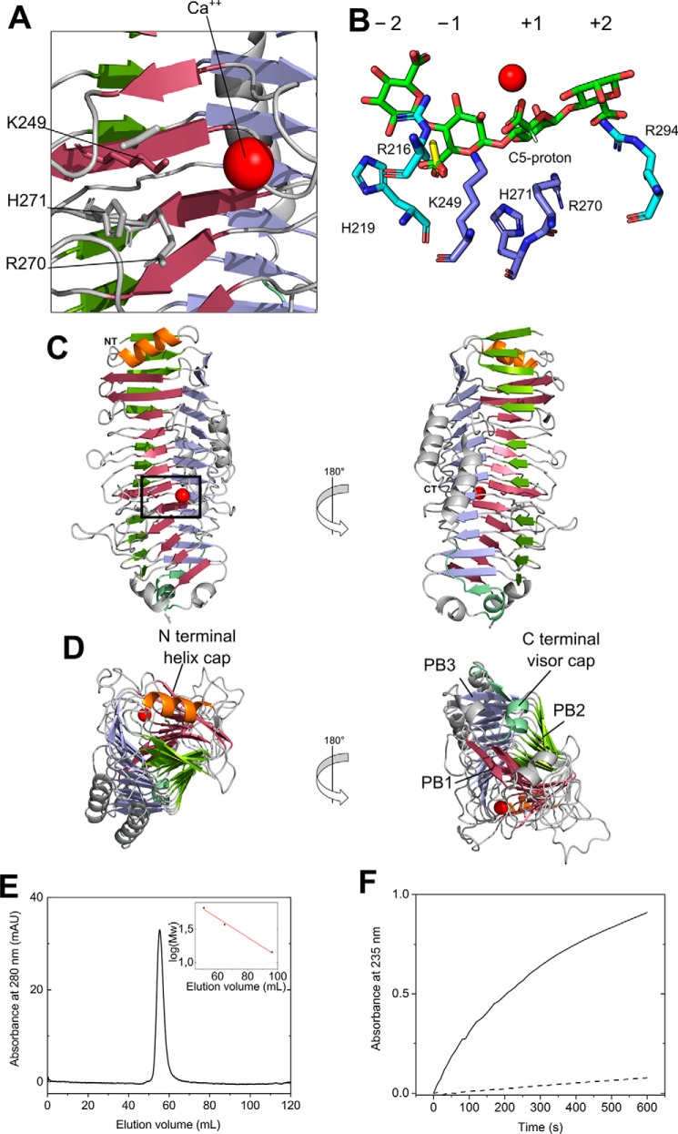
Figure 6.
Ribbon representation of BcelPL6 (PDB 6QPS). A, zoom-in of active site with PL6 conserved catalytic residues Lys-249 and Arg-270 as well as the His-271, situated between subsites −1 and +1, and the neutralizing Ca2+ (red). B, docked DP4M with subsites indicated. The yellow molecule is an acetate found in the crystal structure presumably from the crystallization solvent. C, overall structure of BcelPL6; black box indicates the active-site zoom-in in A. D, N- and C-terminal parts of the β-helix with the capping features and sheets (PB1–PB3) named. E, analytical SEC of BcelPL6 (Superdex 75). The inset is the standard curve of lysozyme, β-lactoglobulin, and BSA yielding a molecular mass of BcelPL6 of 52.3 kDa (theoretical 52.9 kDa). F, increase in absorbance at 235 nm as a function of time of 4 mg ml−1 alginate degradation by 100 nm BcelPL6 dialyzed against 50 mm HEPES, pH 7.3, 150 mm NaCl (solid line), or 50 mm HEPES, pH 7.3, 150 mm NaCl, 1 mm EDTA (dashed line).

