. 2018 Jun 25;35(2):141–154. doi: 10.5511/plantbiotechnology.18.0427a

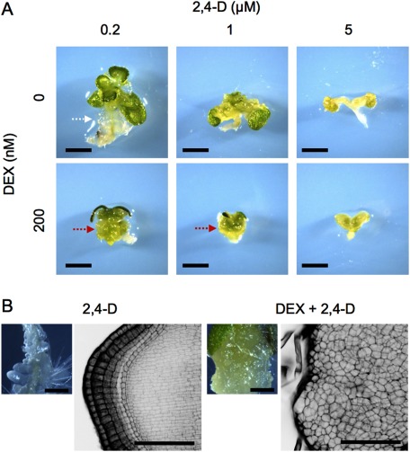
Figure 4. Effects of a simultaneous application of DEX and 2,4-D on Arabidopsis seedling phenotypes. (A) The 35Sp::WOX1-GR plants grown on liquid media containing various concentrations of DEX and/or 2,4-D for three weeks. The relatively low concentration of 2,4-D led to white friable tissue formation (white arrow) in the absence of DEX but green compact calli in the presence of DEX (red arrows). (B) The 35Sp::WOX1-GR plants grown on solid media containing 125 nM DEX and/or 1 µM 2,4-D for 12 day. The left panels are stereomicroscopic images, and the right ones are black-and-white reversal images observed with confocal laser scanning microscope after modified pseudo-Schiff–propidium iodide staining. Scale bars, 2 mm (A), 0.5 mm (B, left panels), 100 µm (B, right panels).

Figure 4. Effects of a simultaneous application of DEX and 2,4-D on Arabidopsis seedling phenotypes. (A) The 35Sp::WOX1-GR plants grown on liquid media containing various concentrations of DEX and/or 2,4-D for three weeks. The relatively low concentration of 2,4-D led to white friable tissue formation (white arrow) in the absence of DEX but green compact calli in the presence of DEX (red arrows). (B) The 35Sp::WOX1-GR plants grown on solid media containing 125 nM DEX and/or 1 µM 2,4-D for 12 day. The left panels are stereomicroscopic images, and the right ones are black-and-white reversal images observed with confocal laser scanning microscope after modified pseudo-Schiff–propidium iodide staining. Scale bars, 2 mm (A), 0.5 mm (B, left panels), 100 µm (B, right panels).