. 2018 Jun 25;35(2):155–159. doi: 10.5511/plantbiotechnology.18.0312a

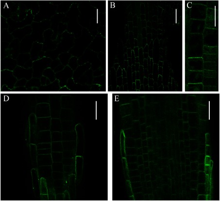
Figure 1. Subcellular localization analysis of eYFP-tagged CVP2 and CVL2 under control of the 35S promoter in Arabidopsis. (A–C) Subcellular localization of eYFP-CVP2 in pavement cells (A), petiole cells (B), and in root cells (C). (D and E) Subcellular localization of eYFP-CVL2 in root tips. Surface view (D) and median longitudinal section (E) of root tips are shown. eYFP-CVP2 and eYFP-CVL2 were excited with 515 nm laser at 30% of laser power and images are taken by Olympus FV1000 confocal laser microscope. Scale bars: 10 µm (A), 30 µm (B), 20 µm (C–E).

Figure 1. Subcellular localization analysis of eYFP-tagged CVP2 and CVL2 under control of the 35S promoter in Arabidopsis. (A–C) Subcellular localization of eYFP-CVP2 in pavement cells (A), petiole cells (B), and in root cells (C). (D and E) Subcellular localization of eYFP-CVL2 in root tips. Surface view (D) and median longitudinal section (E) of root tips are shown. eYFP-CVP2 and eYFP-CVL2 were excited with 515 nm laser at 30% of laser power and images are taken by Olympus FV1000 confocal laser microscope. Scale bars: 10 µm (A), 30 µm (B), 20 µm (C–E).