Figure 2.
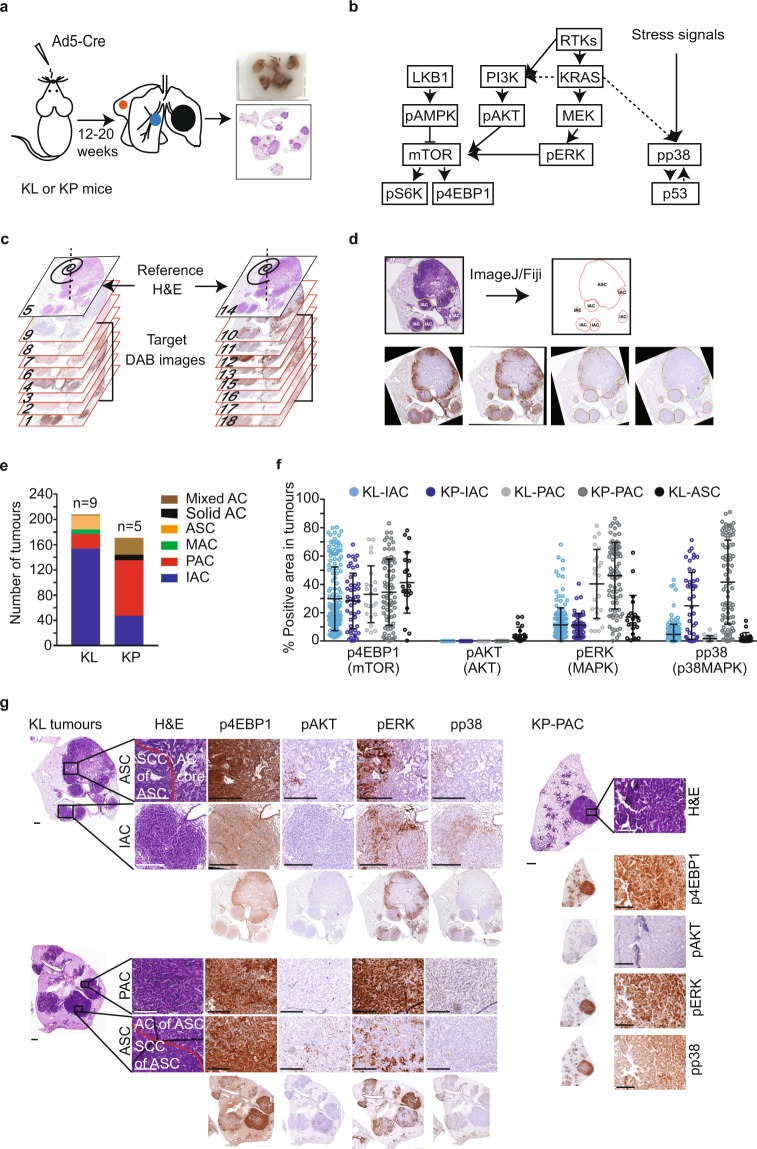
Implementation of Spa-R to analyse oncogenic signalling activities in murine NSCLC histopathologies. (a) Schematic overview depicting FFPE lung tumour tissue block preparation from KrasG12D; Lkb1fl/fl (KL) or KrasG12D; p53fl/fl (KP) GEMMs. Multiple tumours arise in the lung lobes of an individual GEMM. (b) Diagram illustrating oncogenic signalling pathways regulated by KRAS and LKB1 or p53. Solid arrows indicate direct regulation. (c) Schematic overview showing the order of H&E- and DAB-stained tissues sections for image registration. (d) Diagram showing an example image analysis workflow using Spa-R for registration and ImageJ/Fiji for annotation and quantification. The former is used to align DAB images to H&E images, the latter is used to generate annotations and perform ROI-guided quantification of DAB signal on each of the registered images. Red outlines on DAB images are original overlays from H&E-based annotations; green outlines are adjusted overlays due to the distance between tissue sections stained with different antibodies. (e) Graphical representation of the numbers of NSCLC lesions of different histopathologies in 9 KL and 5 KP GEMMs. In mixed ACs (adenocarcinomas) multiple histological features are found in one tumour. (f) Scatterplot showing areas (%) of p4EBP1 (marking mTOR activation), pAKT (marking AKT activation), pERK (marking MAPK activation) and pp38 (marking p38 activation) in individual adenosquamous carcinomas (ASCs), papillary adenocarcinomas (PACs), or invasive adenocarcinomas (IACs). Statistical analysis: one-way ANOVA multiple comparison (Kruskal-Wallis test, nonparametric test) with uncorrected Dunn’s test. (g) Representative images illustrating ASC, PAC, and IAC histological features (H&E staining), as well as the stainings of p4EBP1, pAKT, pERK, and pp38 in a histopathology-specific manner. Scale bars, 500 µm for KL upper panel (entire) images and KL lower panel H&E image and KP H&E image; 100 µm for the others.

