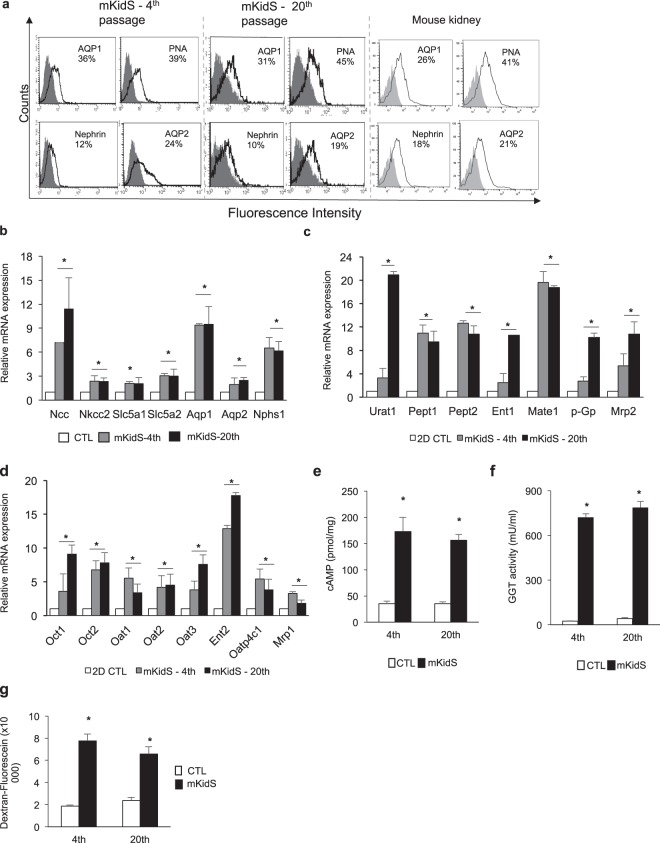
Figure 4.
Reproducibility and maturity of kidney spheroids. (a) FACS analysis of tubule segment markers in mouse kidney spheroid generated by immortalized cells at the 4th and 20th passages. As a positive control, dissociated mouse whole kidney (at 6 weeks of age) was used. Mouse kidney spheroid at the 4th and 20th passages and dissociated whole mouse kidney were stained with anti-AQP1, PNA, nephrin, or AQP2 (open histograms), or isotype control mAb (grey histograms). (b) Quantitative RT-PCR analysis of kidney-specific segment markers in mKidS generated by cells at the 4th and 20th passages compared with 2D controls (2D CTL). n = 8 for 2D CTL and n = 6 for mKidS at the 4th and 20th passages. (c,d) Relative mRNA expression of apical (c) and basolateral (d) transporters in mKidS at the 4th and 20th passages compared with 2D cultured cells. n = 8 for 2D CTL and n = 6 for mKidS at the 4th and 20th passages. (e) Responses to parathyroid hormone (PTH) of mKidS at the 4th and 20th passages compared to 3D CTL. (f) GGT activity in mKidS at the 4th and 20th passages compared to 3D CTL. (g) Alexa Fluor 488-conjugated dextran uptake of mKidS at the 4th and 20th passages compared with 3D CTL. n = 6 for 3D CTL and mKidS at the 4th and 20th passages for E-G. All data are shown as the means ± s.e.m. *p < 0.05 compared with 2D or 3D control by unpaired Student’s t-test.

