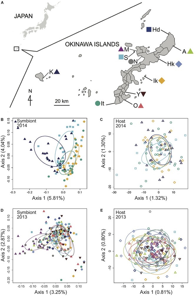
FIGURE 1.

Genomic analysis of the light organ symbionts of Siphamia tubifer reveals structure in populations of symbiotic Photobacterium mandapamensis despite a lack of structure in the host fish. (A) Samples were collected from ten locations in 2013 and from four locations in 2014 in the Okinawa Islands, Japan. Principal coordinates analysis of genetic differentiation of (B) symbiotic P. mandapamensis across 534 loci analyzed from light organs sampled in 2014, (C) the corresponding S. tubifer hosts across 8,637 SNPs, (D) symbiotic P. mandapamensis across 465 loci analyzed from light organs sampled in 2013, and (E) the corresponding S. tubifer hosts across 8,637 SNPs. Points represent individuals along the first and second axes of genetic variation. The first 20 axes of variation for each analysis are depicted in Supplementary Figure S5. Ellipses indicate standard deviation for symbiont populations sampled at each location.
