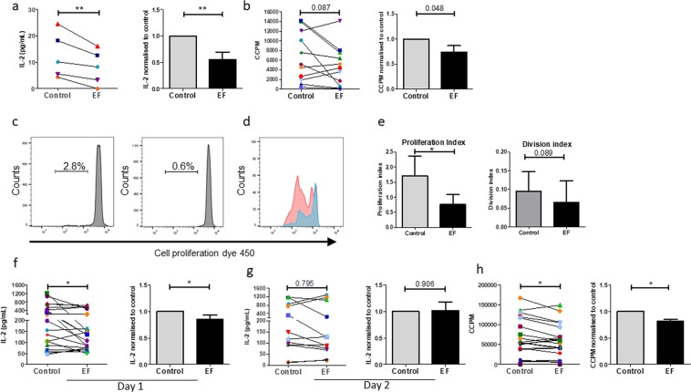
Figure 2.
EF exposure induces changes in PBMC and T cell activation and proliferation. Human PBMCs lymphocytes were activated by PPD recall antigen (5 µg/ml) and LPS (1 µg/ml), with (EF) or without (Control) exposure to EF 150 mV/mm for 4 h. (a) Levels of IL-2 in culture supernatants were determined by ELISA 36 hours following EF-exposure. (b) Cell proliferation was determined on day 5 post EF-exposure by 3H-thymidine incorporation. Human T lymphocytes were stimulated with anti-CD3/CD28 antibodies. (c) Example of proliferation histograms for PPD/LPS stimulated PBMCs without and with EF as assessed by cell proliferation dye, CellTrace Violet. The percentage of reactive cells proliferating are shown. (d) A direct comparison of the mean number of cell divisions by PMBCs activated by PPD/LPS with and without EF exposure. (e) The change in proliferation index (total number of divisions divided by the number of cells that went into division) and division index (mean number of cell divisions that a cell in the original population has undergone) for PPD/LPS activated cells with and without EF exposure (n = 3). Levels of IL-2 in culture supernatants were determined by ELISA (f) 18 hours (day 1) and (g) 36 hours (day2) following EF-exposure. (h) T cell proliferation was determined on day 3 post EF-exposure by 3H-thymidine incorporation. For a, b, f, g and h, data plotted represent individual donor values and right panels represent the normalised data determining the ratio of EF-exposed cells to control cells for each donor; mean values ± SEM. *p < 0.05, **p < 0.01.

