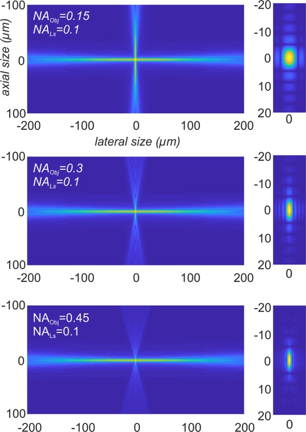
Figure 6.

Modelling the PSF of a light sheet microscope. Simulations of PSFIL and PSFdet of a light sheet microscope for NALS = 0.1 and NAObj = {0.15, 0.3, 0.45} (left). The according effective point spread function PSFLSM is depicted on the right side. For a 2x objective with NAObj = 0.15 the effect of the detection PSF is small, since most outer parts of PSFdet are multiplied with values that are close to zero. However, the higher NAObj becomes, the more it effects the shape of PSFLSM. To enhance the visibility of the side lobes of the PSFs, the images were gamma corrected by γ = 0.4.
