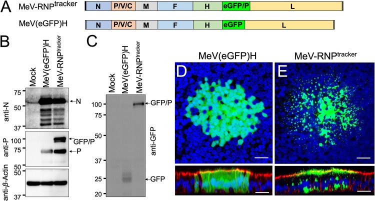
FIG 4.
Generation and characterization of MeV-RNPtracker. (A) Schematics of the MeV-RNPtracker genome (top) and of the MeV(GFP)H genome (bottom) control virus. In MeV-RNPtracker, GFP was fused in frame with a second copy of the P protein (GFP/P), and the transcription unit was inserted between the H and L genes. In MeV(GFP)H, a transcription unit expressing GFP was inserted in the same position. (B and C) Immunoblot characterization of the P proteins expressed by the two viruses. In panel B, the expression of the N (top), P and GFP/P (center), or control actin (bottom) proteins was analyzed. In panel C, the expression of GFP/P and GFP proteins were analyzed. At 3 days postinfection, the GFP expression of MeV(GFP)H (D) and MeV-RNPtracker (E) was examined by confocal microscopy. Nuclei were stained with DAPI (blue), and F-actin was visualized with phalloidin (red, only shown in the bottom panel vertical sections). Images are representative from n = 6 samples (two technical replicates from three human donors [biological replicates]). Scale bars, 20 μm.

