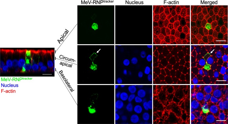
FIG 6.
In newly infected cells, MeV RNPs localize to the circum-apical network. Cells were infected with MeV-RNPtracker, fixed, and imaged at 36 hpi by confocal microscopy. Cells were counterstained for F-actin with phalloidin (red), and nuclei were visualized with DAPI (blue). The left panel is a vertical section view, and the vertical bars indicate the plane of view for the series of en face images on the right. The right panels show maximum intensity projection images of three to five z-stacks at the indicated apical, circumapical, and basolateral regions. White arrows indicate MeV RNPs along the circumapical region of the F-actin network in newly infected cells. Images are representative from n = 6 samples (two technical replicates from three human donors [biological replicates]). Scale bars, 10 μm.

