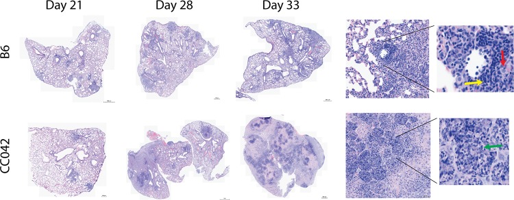
FIG 2.
Changes in lung pathology during M. tuberculosis infection. Lung lobes obtained from B6 and CC042 mice at days 21, 28, and 33 postinfection were stained with hematoxylin and eosin. Images are representative of those from 6 mice per strain per time point. (Insets) Magnified images from day 33 show lymphocytes (yellow arrow), macrophages (red arrow), and neutrophils (green arrow).

