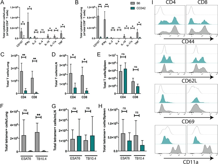
FIG 6.
CC042 mice have a defect in T cell recruitment to the lung and lack CD11a expression. (A, B) Intracellular cytokine staining (ICS) of CD4 (A) and CD8 (B) T cells reveals a defect in the number of cytokine-producing T cells in the lungs of CC042 mice. (C to E) Total number of CD4 and CD8 T cells in the lung (C), mediastinal lymph node (D), and spleen (E). (F to H) Total number of ESAT-6 (CD4) or TB10.4 (CD8) tetramer-positive cells in the lung (F), mediastinal lymph node (G), and spleen (H). Bar plots show the mean + SD. Welch’s t test was used to determine significance. *, P < 0.05; **, P < 0.01; ns, not significant. (I) Histograms of CD4 (left) and CD8 (right) T cells stained for activation and migration markers CD44, CD62L, CD69, and CD11a for the isotype control (top, light gray trace), CC042 mice (middle, teal trace), and B6 mice (bottom, gray trace).

