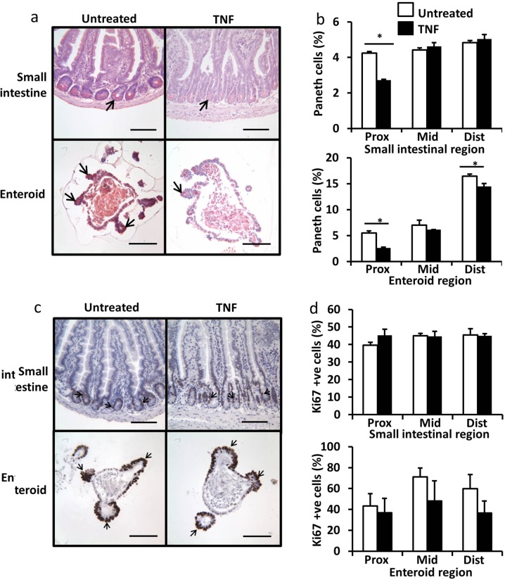
Fig. 2. TNF causes Paneth cell degranulation in proximal SI but does not change the rate of cell proliferation.
Proximal SI and enteroids stained with Sirius red (a). Percentage of Sirius red stained Paneth cells in untreated (white) and TNF treated (black) proximal, middle and distal derived SI (top) and enteroids (bottom) (b). Proximal SI and enteroids stained with Ki67 (c). Percentage of Ki67 stained epithelial cells in untreated (white) and TNF treated (black) proximal, middle and distal derived SI (top) and enteroids (bottom) (d). For animal study quantified from n = 6 mice per group, for enteroid study n = 6, N = 3, *p < 0.05. Scale bars = 100 µm.

