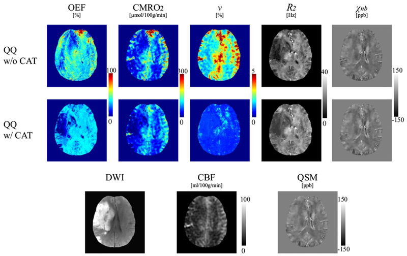
Figure 4.

Comparison of OEF, CMRO2, v, R2 and χnb maps between QQ without and with CAT in a stroke patient imaged 6 days post stroke onset. In the CMRO2 and OEF maps, the lesion can be distinguished more clearly with QQ with CAT. For QQ with CAT, a low OEF region is clearly visualized and contained with the lesion region as defined on DWI, but a low OEF region obtained with QQ without CAT is not as well localized nor contained within the lesion as defined on DWI. QQ with CAT generally shows lower v in the DWI-defined lesion. The contrast in v in QQ without CAT result is similar in appearance to that of CBF. QQ with CAT shows generally higher R2 and χnb maps.
