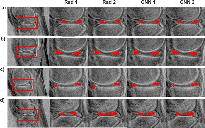
Figure 6.
Examples illustrating the results of the manual and automatic segmentations. a) and b) show results for which a large level of agreement was obtained for each method. In c) the second radiologist, in comparison to the first one, outlined the right part of meniscus differently, but both deep models pointed out this part of the image as corresponding to the meniscus. For the images presented in d) the first radiologist outlined the meniscus in a conservative manner. In comparison, the second radiologists and both CNNs generated larger ROIs.

