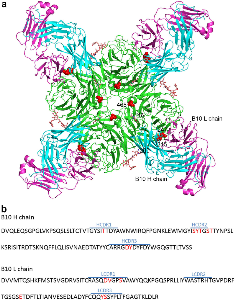
Fig. 4. Crystal structure of the Fab of mAb B10 in complex with the NA of A(H3N2) Virus.
Antibody B10 was raised against the NA of A/Minnesota/11/2010 (MN/10), and because it reacted with the NA of A(H3N2) virus until the acquisition of NA245 glycosylation site, it was selected in our study to explore the structural basis underlying the impact of this glycosylation site on antibody binding to NA. a, Overall structure of the antibody–antigen complex, showing four B10 Fabs (H chain in cyan, L chain in magenta) bind to an NA tetramer; residues 245, 247, and 468 are highlighted in red, and glycans at residues 146 and 200 are shown as sticks. b, Amino acid sequence of the B10 Fab variable regions. CDR regions of the H and L chains were defined using IMGT/V-QUEST50, and residues that are in contact with NA in the complex structure were highlighted in red.

