Fig. 7:
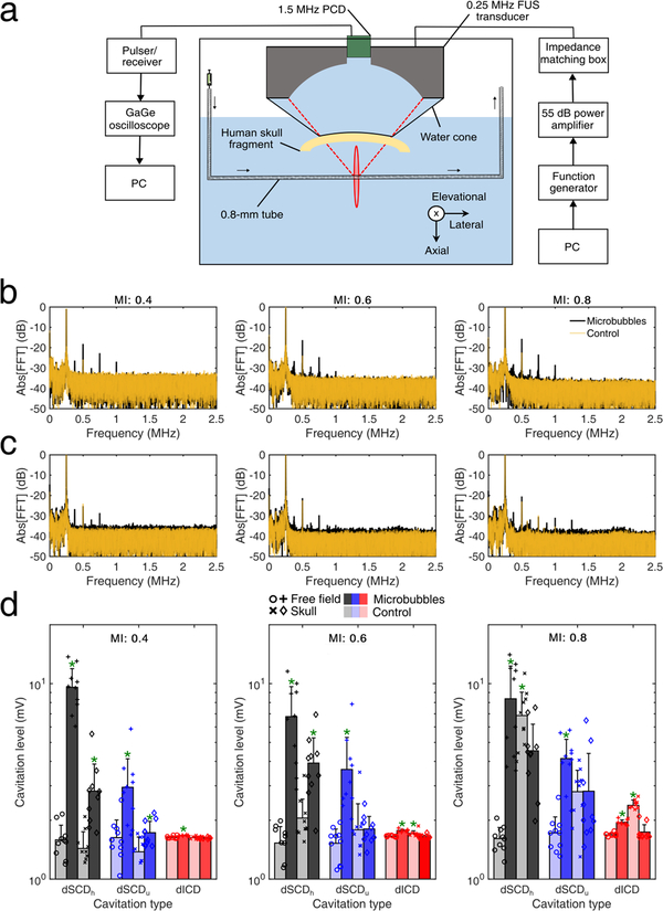
Passive cavitation detection through the human skull. (a) In vitro setup for passive cavitation detection. A 0.8-mm tube filled with Definity microbubbles was used as a vessel-mimicking phantom. (b) Spectra of control (transparent orange line) and microbubble (black line) acoustic emissions for mechanical index (MI) of 0.4 (left), 0.6 (middle), and 0.8 (right) in free field. (c) Spectra of control and microbubble acoustic emissions through the human skull. (d) Cavitation levels in free-field (circles, plus signs) and through the human skull (crosses, diamonds), for control (light bars) and microbubbles (dark bars), at MI of 0.4 (left), 0.6 (middle), and 0.8 (right). Data presented as mean ± standard deviation (n=10 pulses). *: p < 0.05.

