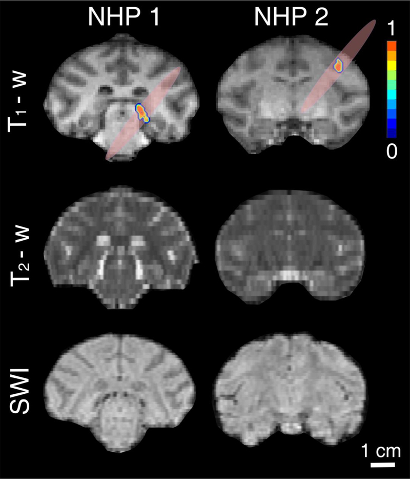
Fig. 9:
In vivo feasibility in a non-human primate (NHP) model. Coronal T1-weighted, T2-weighted and susceptibility-weighted imaging (SWI) for NHPs 1 (left) and 2 (right). T1-weighted MRI confirmed blood-brain barrier opening in the thalamus (NHP 1) and dorsolateral prefrontal cortex (NHP 2), using the clinical focused ultrasound (FUS) transducer with clinically relevant parameters (MI: 0.4) and FDA-approved Definity microbubble dose (10 μl/kg). T2-weighted and SWI showed that there is no acute hemorrhage or edema after the FUS treatment. Color bar: normalized contrast enhancement. Scale bar: 1 cm.

