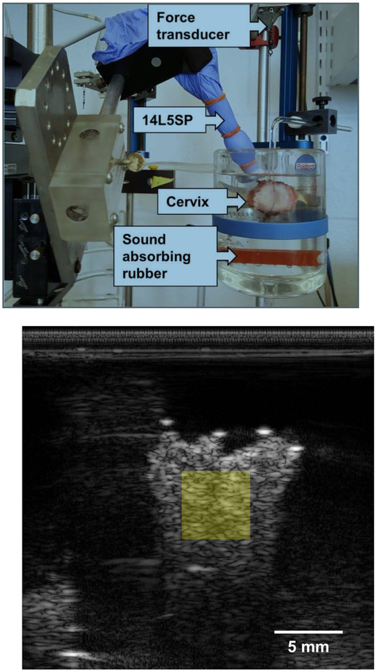
Figure 1:
(a) A representative photograph of the experimental setup for measuring muscle force generation and RF data acquisition. The cervix specimen, the 14L5SP ultrasound transducer, and the force transducer are labeled with arrows. Two sutures are shown that are inserted through the endocervical canal. One of those sutures attaches the cervix to the bottom of the organ bath. The other suture extends up to the force transducer. Orange inserts in the organ bath are sound absorbing rubber, helping to minimize clutter in the (b) B-mode image. The semitransparent, yellow square represents where in the sample backscatter parameters are estimated.

