Fig. 6.
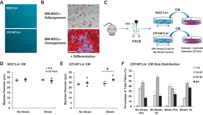
Muscle-derived CD146+Lin− pericytes secrete factors in response to in vitro mechanical strain that positively influence myotube growth. Neuron-glial antigen 2-positive lineage-negative (NG2+Lin−) and CD146+Lin− pericytes were isolated from unstimulated mouse muscle by fluorescence-activated cell sorting. A: morphology of pericytes after 1 wk in culture. Whereas NG2+Lin− pericytes demonstrate a punctate shape, CD146+Lin− pericytes exhibit a fibroblast-like morphology. B: pericytes and bone marrow-derived MSCs (BM-MSCs) were plated on plastic culture dishes and evaluated for differentiation capacity. Pericytes did not demonstrate capacity for myogenesis, adipogenesis, or osteogenesis (not shown). Control BM-MSCs display capacity for adipogenesis based on Oil Red O Staining (top) and osteogenesis based on Alizarin Red S staining (bottom); n = 3. C: muscle-resident pericytes were subjected to a single bout of in vitro mechanical strain using a Flexcell System (laminin-coated plates, 10% strain, 1 Hz, 1 h). FACS, fluorescence-activated cell sorting. D and E: 24 h following in vitro mechanical strain, pericyte-derived conditioned media (CM; strained or no strain control) was collected and subsequently added to mature myotubes in culture. Whereas addition of NG2+Lin− CM did not influence myotube growth (D), CD146+Lin− CM significantly increased myotube growth (E) at l day (1D) postincubation. F: myotube size distribution is provided for cells exposed to CD146+Lin− conditioned media. ^ Time main effect, P < 0.05; #treatment main effect, P < 0.05. Values are means ± SE (n = 4).

