Fig. 7.
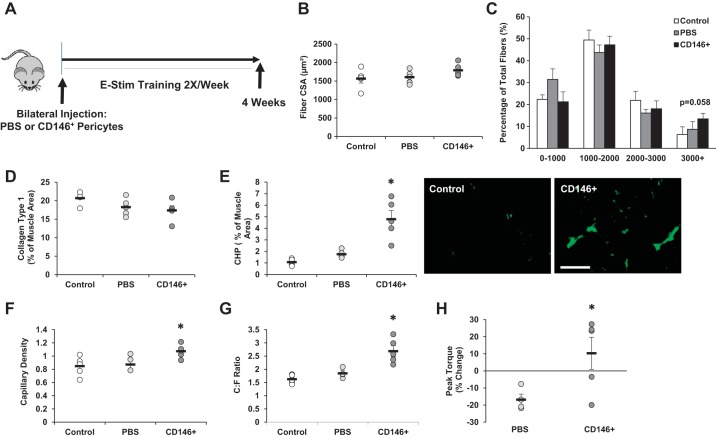
Muscle-derived CD146+Lin− pericytes contribute to skeletal muscle remodeling in response to training. A: experimental design for CD146+ pericyte transplantation and training study. CD146+Lin− pericytes were isolated from unstimulated mouse hindlimb muscle by fluorescence-activated cell sorting. Pericytes were bilaterally injected into tibialis anterior (TA) and gastrocnemius muscles of 16-wk-old mice (“CD146+”). A separate group of mice were injected with PBS as a control (“PBS”). A third group of mice received no treatment [“Control”; no injection, no electrical stimulation (e-stim)]. Pericyte and PBS-injected mice were exposed to unilateral e-stim immediately postinjection. E-stim treatment was applied 2 days/wk, for a total of 4 wk (8 sessions total). Twenty-four hours following the final stimulation, muscles were harvested and prepared for immunofluorescence analysis. B–H: the following analyses are reported for the TA muscle: mean fiber cross-sectional area (CSA; B), minimum 200 fibers analyzed per section, fiber size distribution (C), collagen type 1 content (D), collagen degradation based on collagen hybridizing peptide (CHP) assay (E), capillary density (F), capillary to fiber ratio (C:F; G), and change in peak torque (H) from beginning to end of study. Values are means ± SE (n = 6). *P < 0.05 vs. Control and PBS.

