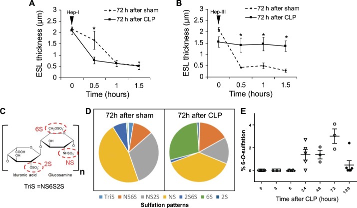
Fig. 2.
The postseptic, reconstituted endothelial glycocalyx is remodeled. A: at the 72-h time point characterized by pulmonary endothelial heparan sulfate (HS) reconstitution, we injected 1 unit of heparinase I (Hep I) into the jugular vein, which degraded both sham and post-cecal ligation and puncture (CLP) pulmonary endothelial glycocalyx heparan sulfate. B: in contrast, 1 unit of heparinase III (Hep III) into the jugular vein did not degrade postseptic pulmonary endothelial heparan sulfate. C: major sulfation positions on heparan sulfate constituent disaccharides. D: postseptic, reconstituted pulmonary endothelial heparan sulfate had a significant increase (P < 0.05) in disaccharides with 6-O-sulfation (percentage of total sulfated disaccharides) as compared with heparan sulfate from sham animals. E: the percentage of 6-O-sulfated disaccharides increased over time in the plasma of post-CLP mice. ESL, endothelial surface layer. *P < 0.05 by Student’s t test; n = 3–5 per group.

