Fig. 5.
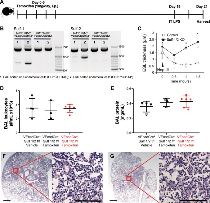
Loss of sulfatase-1 (Sulf-1) is not sufficient to cause impaired inflammation in nonseptic animals. A: Sulf-1f/f Sulf-2f/f VEcadCreERT2+ or – animals received tamoxifen or vehicle control injections intraperitoneally for 5 consecutive days (1 mg/day). Recombination of genes and pulmonary endothelial glycocalyx characteristics were evaluated 2 wk after the last injection of tamoxifen (or vehicle). Knockout or control mice were alternatively challenged with intratracheal (IT) LPS (3 μg/g body wt) at the same time point. B: we confirmed cell-specific, inducible recombination of Sulf-1 and Sulf-2 with DNA gels. Lane 1: DNA from pulmonary nonendothelial cells (CD31–/CD144–). Lane 2: DNA from pulmonary endothelial cells (CD31/C+D144+). C: pulmonary endothelial glycocalyx of Sulf-1/2 knockout animals was resistant to heparinase III (Hep III) degradation, similar to the postseptic endothelial glycocalyx resistance to heparinase III observed in wild-type mice (Fig. 2B). Control animals used were Sulf-1f/f Sulf-2f/f VEcadCreERT2– (floxed gene alone without Cre recombinase), treated with tamoxifen. D: number of leukocytes in bronchoalveolar lavage (BAL) fluid in Sulf-1/2 knockout animals did not differ from control groups 2 days after IT LPS. E: protein concentration of BAL fluid was similarly not different among the experimental groups. F: lung histological section from a control animal (Sulf-1f/f Sulf-2f/f VEcadCreERT2–, treated with tamoxifen) had clear consolidation. G: lung histological section from a Sulf-1/2 knockout animal similarly had evidence of lung inflammation. Scale bars on lower-magnification images = 500 μm and those on higher-magnification images = 100 μm. *P < 0.05 by one-way ANOVA with post hoc Tukey test; n = 3–6 each group.

