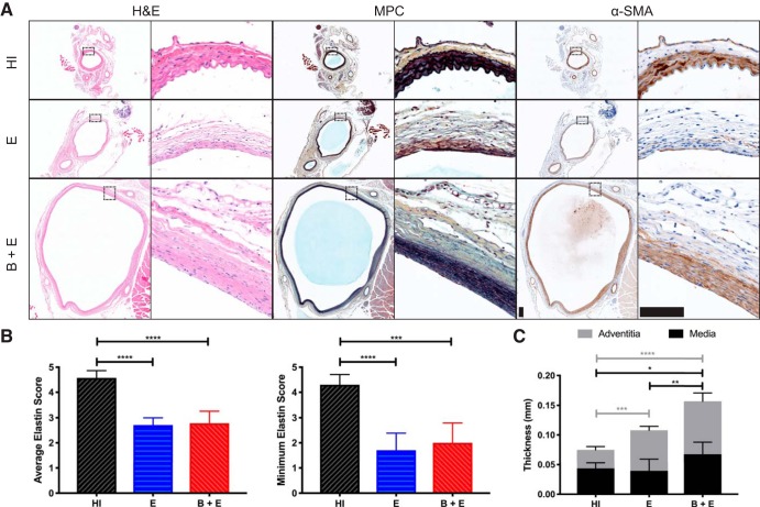
Fig. 5.
Histology and immunohistochemistry (IHC). A: hemotoxin and eosin (H & E), Movat’s Pentachrome (MPC), and α-smooth muscle actin (α-SMA) staining at ×4 (left) and ×40 (right) magnifications. Scale bar, 0.1 mm for all images. B: results of semiquantitative scoring analysis, where 5 = healthy elastin sheets present, 3 = degraded elastin fragments present, and 1 = elastin not present. Averaging all 4 quadrants (left) and using the minimum quadrant score (right) both reveal decreased elastin content in elastase-only (E) and β-aminopropionitrile-elastase (B + E) groups (P < 0.001). C: aortic wall thickness from histology, as defined by medial and adventitial layers. *P ≤ 0.05; **P ≤ 0.01; ***P ≤ 0.001; ****P ≤ 0.0001. HI, heat inactivated.

