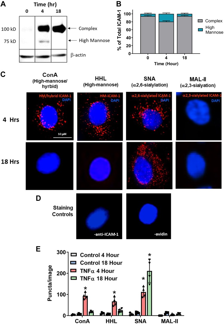
Fig. 2.
TNF-α forms endothelial high-mannose (HM)-intercellular adhesion molecule-1 (ICAM-1) in a time-dependent manner. Human umbilical vein endothelial cells (HUVECs) were treated with 10 ng/mL TNF-α for 0, 4, or 18 h, and either lysates were collected for Western blot analyses or cells were processed for proximity-ligation assay (PLA). A: representative Western blot for ICAM-1 expression. The 100-kDa band represents the fully glycosylated complex ICAM-1, and the 75-kDa band represents the hypoglycosylated high-mannose ICAM-1. Kif, Kifunensine; Swain, swainsonine. B: quantification of HM-ICAM-1 as a percentage of total ICAM-1. Data are means ± SE, n = 3. C: HUVECs were treated as above and subject to a PLA for HM/hybrid, HM, α2,6-sialylated, and α2,3-sialylated ICAM-1. Shown are representative images from each time point. Red puncta represents positive PLA signal, blue staining is DAPI nuclear stain. ConA, concanavalin A; HHL, Hippeastrum hybrid amaryllis; SNA, Sambucus nigra; MAL-II, maackia amurensis lectin II. D: PLA staining controls where one PLA reagent was left out (left, no anti-ICAM-1; right, no avidin). E: quantification of PLA puncta. For each replicate, puncta were counted in three fields and averaged. Each symbol represents an independent replicate. Data are means ± SE, n = 4. *P ≤ 0.05 compared with respective time control by 1-way ANOVA with Tukey’s posttest.

