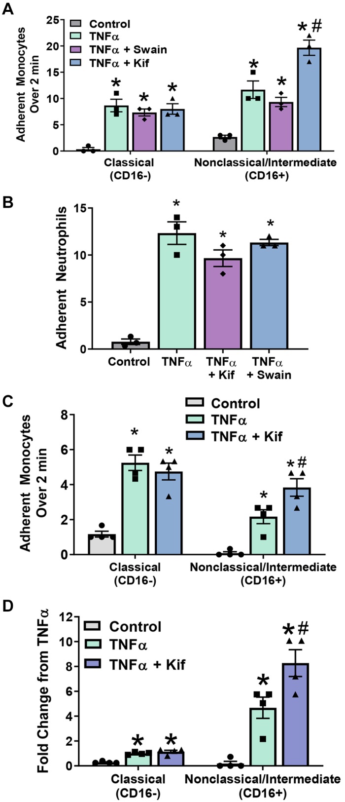
Fig. 4.

CD16+ monocyte adhesion is enhanced by endothelial high-mannose epitopes. A: human umbilical vein endothelial cells (HUVECs) were treated as above and exposed under flow to fluorescently labeled CD16+ and CD16− monocytes. Each symbol represents the average of an independent experiment. Kif, kifunensine; Swain, swainsonine. Data are means ± SE, n = 3. P ≤ 0.05 compared with control (*) or TNF-α (#) by 1-way ANOVA with Tukey’s posttest within monocyte groups. B: HUVECs were treated, and neutrophil adhesion under flow was determined as described. Data are means ± SE, n = 3. *P ≤ 0.05 compared with control alone by 1-way ANOVA. C: monocyte adhesion under flow to HUVECs at physiological ratios (225,000 cells/mL CD16− and 25,000 cells/mL CD16+). Each bar represents the average of 4 independent experiments. Data are means ± SE, n = 4. P ≤ 0.05 compared with control (*) or TNF-α (#) by 1-way ANOVA with Tukey’s posttest within monocyte groups. D: monocyte adhesion from C as a fold change compared with CD16− adhesion to TNF-α-treated HUVECs. Each symbol represents the average of 4 independent experiments. Data are means ± SE, n = 4. P ≤ 0.05 compared with control (*) or TNF-α (#) by 1-way ANOVA with Tukey’s posttest within monocyte groups.
