Fig. 2.
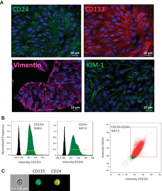
Characterization of scattered tubular-like cells (STCs) isolated from pig kidneys. A: representative immunofluorescence staining (original magnification: ×40) for the STC surface markers CD24 (green), CD133 (red), vimentin (pink), and kidney injury molecule (KIM)-1 (green) in isolated swine STCs. B: flow cytometry analysis of isolated STCs showing that 98.6% of cells expressed CD133, 97.0% expressed CD24, and 97.5% were double positive for CD133 and CD24. C: representative image of a CD133+ (green)/CD24+ (yellow) cell.

