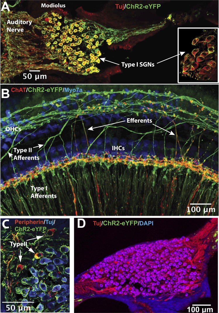
Fig. 2.
Expression pattern of ChR2R-eYFP in the cochlea. A: cross section through the modiolus from a Bhlhb5-ChR2R heterozygous mouse cochlea showing ChR2R-eYFP (anti-GFP in green) expressed in almost all type I spiral ganglion neurons (SGNs; labeled by class III β tubulin antibody, TuJ in red). Yellow shows double labeling. Inset, with similar immunostaining, shows a high-power view of type I SGN cell bodies from a whole-mount of a Bhlhb5-ChR2R homozygous cochlea. B: whole-mount of a portion of the organ of Corti. ChR2R-eYFP (in green) is strongly expressed in radially oriented type I afferent fibers and spirally oriented type II afferent fibers. Expression was also observed in olivocochlear fibers (“Efferents”) near inner hair cells (IHCs), which were labeled by anti-choline acetyltransferase (ChAT, in red). Inner and outer (OHC) hair cells did not show expression (labeled blue by Myo7a antibody). C: cross section of a modiolus from a Bhlhb5-ChR2R heterozygous cochlea showing ChR2R-eYFP (in green) expressed in type I and type II SGNs. Type II SGNs were identified by anti-peripherin staining (red). D: cross section through the modiolus from a control mouse (ChR2R-eYFP+/fl). No ChR2R-eYFP (green) was found in SGNs (TuJ antibody, in red) in these control mice.

