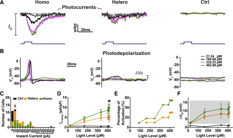
Fig. 3.
Expression-level dependent optogenetic excitation of spiral ganglion neurons (SGNs) in whole cell recordings. Photocurrents (IO; A) and photodepolarization (∆VO; B) were observed in SGNs from homozygous (Homo), heterozygous (Hetero), and control (Ctrl) littermates at postnatal day (P)2 to P4. Light pulses (10 ms; blue traces) were presented to cells at a series of power levels. Dashed lines are representative baselines at the maximum light power level used (413 µW). Vm, membrane potential. C: histogram of photocurrent magnitude distribution at maximum light stimulation. Bin size = 10 pA. The cells included here met the criterion (red arrowhead) of IO ≥ 10 pA. D–F: averaged photocurrent density (IO-density), percentage of SGNs with optically evoked action potentials, and subthreshold photodepolarization (∆VO) as a function of light power level are derived from 15 homozygous, 12 heterozygous, and 8 control cells. Significance was examined by the Mann–Whitney t test. *P < 0.05.

