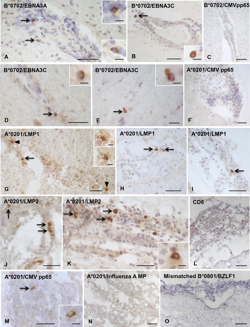
FIG 4.
Localization of CD8 T cells specific for EBV latent proteins in MS brain sections. Cells binding pentamers coupled with peptides from different EBV latent proteins were visualized in bright field in brain sections from HLA-A*0201+, B*0702+, and B*0801+ MS donors. (A to C, donor MS356, B*0702+) Presence of CD8 T cells specific for B*0702-restricted EBNA3A (A) and EBNA3C (B) peptides, but not for B*0702-restricted CMV pp65 peptide (C), in the inflamed meninges; the insets show the cells indicated by the arrows in panels A and B at high-power magnification. (D to F, donor MS289, A*0201+/B*0702+) Perivascular CD8 T cells specific for B*0702-restricted EBNA3C peptide are indicated by the arrows in panels D and E and shown at high-power magnification in the insets. (F) Absence of CD8 T cells specific for A*0201-restricted CMV pp65 peptide in a meningeal infiltrate. (G to O, donor MS79, A*0201+) Presence of CD8 T cells specific for A*0201-restricted LMP1 peptide (G to I, arrows; the insets in panel G show the cells indicated by the arrowheads at high-power magnification) and LMP2 peptide (J and K, arrows) in the meninges where numerous scattered CD8 T cells are also detected (L). In adjacent sections, one CD8 T cell specific for A*0201-restricted CMV pp65 peptide (M; the inset shows the cell indicated by the arrow at high-power magnification) but no CD8 T cells specific for A*0201-restricted influenza A MP peptide (N) or cells binding B*0801/BZLF1 pentamer (O) were detected. Nuclei were stained with hematoxylin. Bars, 100 μm (C, L, and O), 50 μm (A, B, D, F to K, M, and N), 20 μm (E and inset in panel M), and 10 μm (insets in panels A, B, D, E, G, and K).

