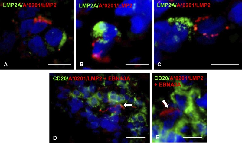
FIG 9.
EBV-specific CD8 T cells contact EBV-infected cells in the MS brain. (A to C) Double immunofluorescence staining of brain sections from the A*0201+ MS79 donor with MAb specific for the EBV latent protein LMP2A (green) and pooled A*0201 pentamers coupled with LMP2 426–434 and LMP2 356–364 peptides (red) reveals the presence of tight contacts between LMP2A-expressing cells and pentamer-binding cells in the meninges. (D and E) Double immunofluorescence staining of a brain section from the same donor with anti-CD20 MAb (green) and pooled A*0201 pentamers coupled with LMP2 426–434, LMP2 356–364, and EBNA3C peptides (red) shows individual pentamer-binding cells in contact with B cells within a B-cell-rich meningeal infiltrate. Nuclei were stained with DAPI. Bars, 20 μm (D) and 10 μm (A to C and E).

