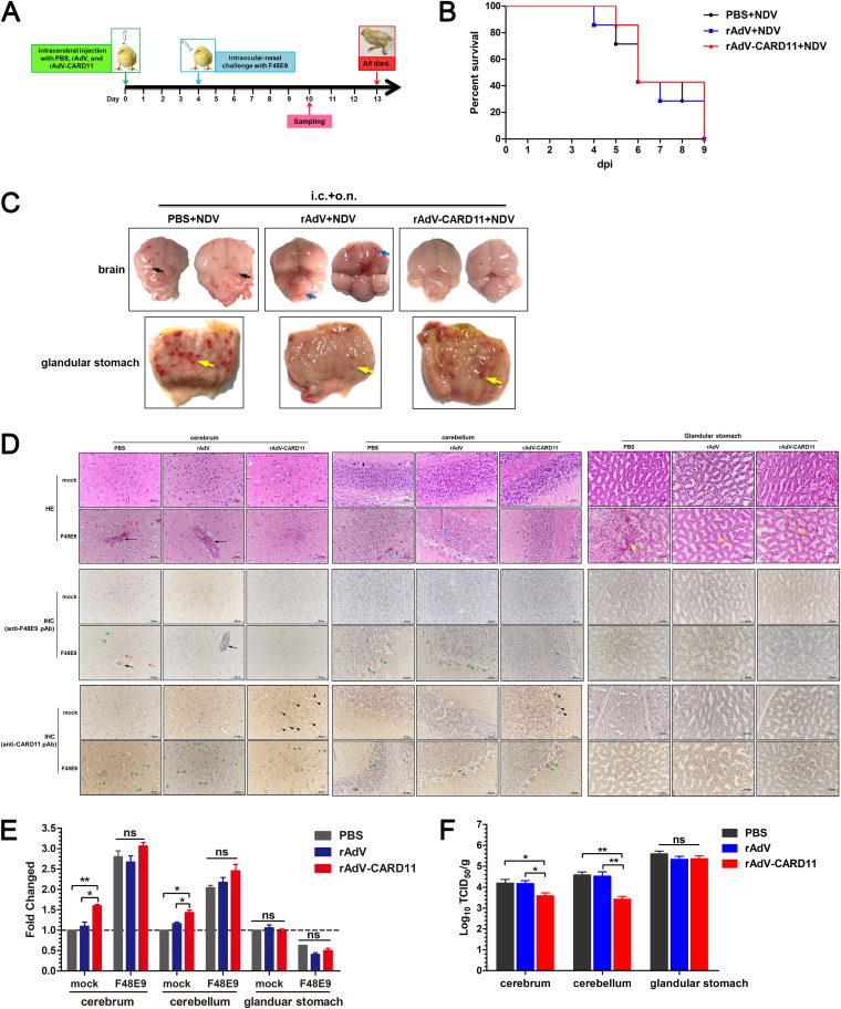
FIG 13.
Intracerebrally overexpressed CARD11 reduces neuropathology and viral replication in the brain. (A) Schematic representation of animal experiments. One-day-old chicks were intracerebrally injected with rAdV-CARD11, rAdV, and PBS. After 4 days, the birds were challenged with F48E9. Tissue samples were collected on day 10. (B) Survival rates of the F48E9-challenged chicks. (C) Lesions on the cerebrum, cerebellum, and glandular stomach at 6 dpc. The black arrows indicate the indistinct boundary between the cerebrum and cerebellum. The blue arrows indicate hyperemia in F48E9-infected brains. The yellow arrows indicate the glandular papillary hemorrhage. i.c., intracerebral injection; o.n., ocular-nasal route. (D) HE and IHC assays at 6 dpc. The black arrows indicate perivascular cuffing in the cerebrum. The red arrows indicate edema in the cerebrum. The blue arrows indicate hemorrhage in the substantia alba of the cerebellum. The yellow arrows indicate the epithelial hemorrhage, swelling, necrosis, and even exfoliation in the compound tubular glands of glandular stomach. The blue arrowhead indicates the detachment of Purkinje cells from the stratum granulosum in the cerebellum. The black arrowheads indicate the detected CARD11 in cells of mock chicks. The green arrowheads indicate the detected virus or CARD11 in the cells of F48E9-challenged chicks. (E) CARD11 expression in the cerebrum, cerebellum, and glandular stomach before and after the F48E9 challenge. (F) The viral replication of F48E9 in the cerebrum, cerebellum, and glandular stomach. The tissues were homogenized, and viral titers in the supernatants were measured in DF-1 cells using the TCID50 method. Representative data from three independent experiments, shown as the means ± SDs (n = 3), were analyzed by two-tailed Student's t tests. ns, not significant; *, P < 0.05; **, P < 0.01.

