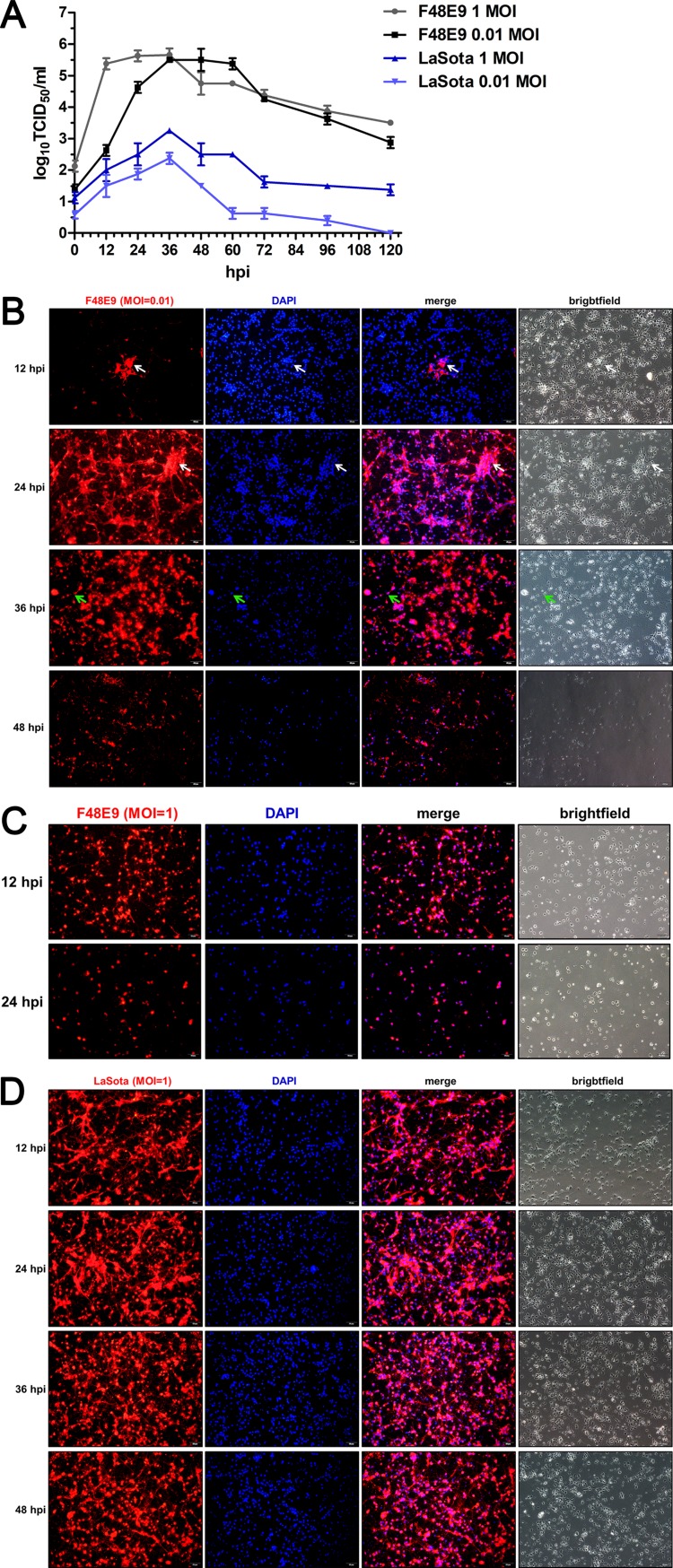
FIG 3.
Virulent NDV shows better replication than lentogenic NDV in chPNCs. (A) Replication of NDV in chPNCs. The cells were infected with F48E9 (MOI = 0.01 or 1) and LaSota (MOI = 0.01 or 1) for 1 h at 37°C. The viral replicates of the culture supernatants at different times after infection were titrated in DF-1 cells. The results are presented as the means ± SDs from three independent experiments. (B to D) The infection of NDV strains in the chPNCs. chPNCs were infected with F48E9 (MOI = 0.01) (B), F48E9 (MOI = 1) (C), and LaSota (MOI = 1) (D) at 12, 24, 36, and 48 hpi. The cells were examined by immunofluorescence assay (IFA) using an anti-NDV mouse PAb (1:200). The white arrows indicate syncytia, and the green arrows indicate axon disruption. Bars, 50 μm.

