FIG 8.
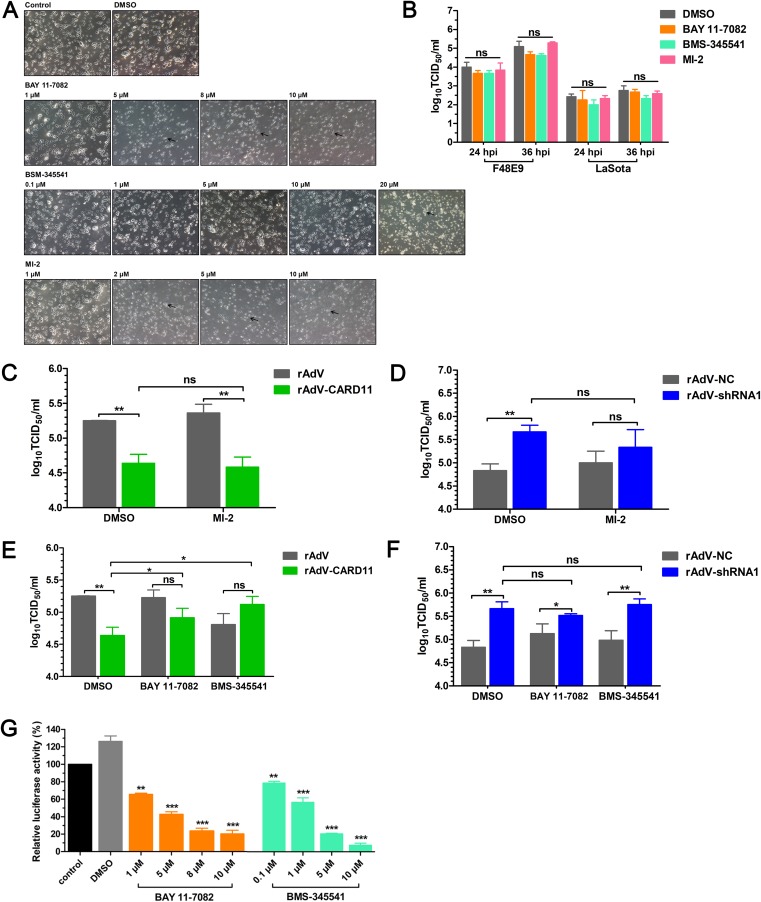
The CBM signalosome plays no role in inhibiting NDV replication in chPNCs. (A) The CPEs of inhibitors in chPNCs. The chPNCs in 12-well plates at day 3 were incubated with different concentrations of inhibitors for 24 h. The treated chPNCs were observed after 24 h. The black arrows indicate axon disappearance and cell body disruption. Bars, 50 μm. (B) NDV replication in inhibitor-treated chPNCs. The cells at day 3 were treated with BAY 11-7082 (1 μM), BMS-345541 (10 μM), MI-2 (1 μM), and DMSO for 24 h, and then the treated chPNCs were infected with the F48E9 strain (MOI = 0.01). The culture supernatants were harvested at 24 and 36 hpi. (C to F) F48E9 replication in CARD11-overexpressing or knockdown chPNCs treated with inhibitors. The cells at day 3 were infected with rAdV, rAdV-CARD11, rAdV-NC, and rAdV-shRNA1 at the same MOI (MOI = 100). At 48 hpi, the cells were treated with MI-2 (1 μM), BAY 11-7082 (1 μM), BMS-345541 (10 μM), and DMSO for 24 h. Then, the treated chPNCs were infected with the F48E9 strain (MOI = 0.01). The culture supernatants were harvested at 36 hpi for titration of F48E9. The viral titers in the supernatants of chPNCs were analyzed via the TCID50 method in DF-1 cells. (G) The inhibitory role of BAY 11-7082 and BMS-345541 in DF-1 cells. The DF-1 cells in 12-well plates were cotransfected with pchNF-κB-TA-luc and pRL-SV40-N at a ratio of 10:1. Twenty-four hours later, the cells were incubated with 1, 5, 8, or 10 μM BAY 11-7082, 0.1, 1, 5, or 10 μM BMS-345541, or DMSO for 24 h and were lysed to quantify the luciferase activity. Renilla luciferase expressed by pRL-SV40-N was used as the normalizing standard. All the representative data, shown as the means ± SDs (n = 3), were analyzed by two-tailed Student's t tests. ns, not significant; *, P < 0.05; **, P < 0.01; ***, P < 0.001.

