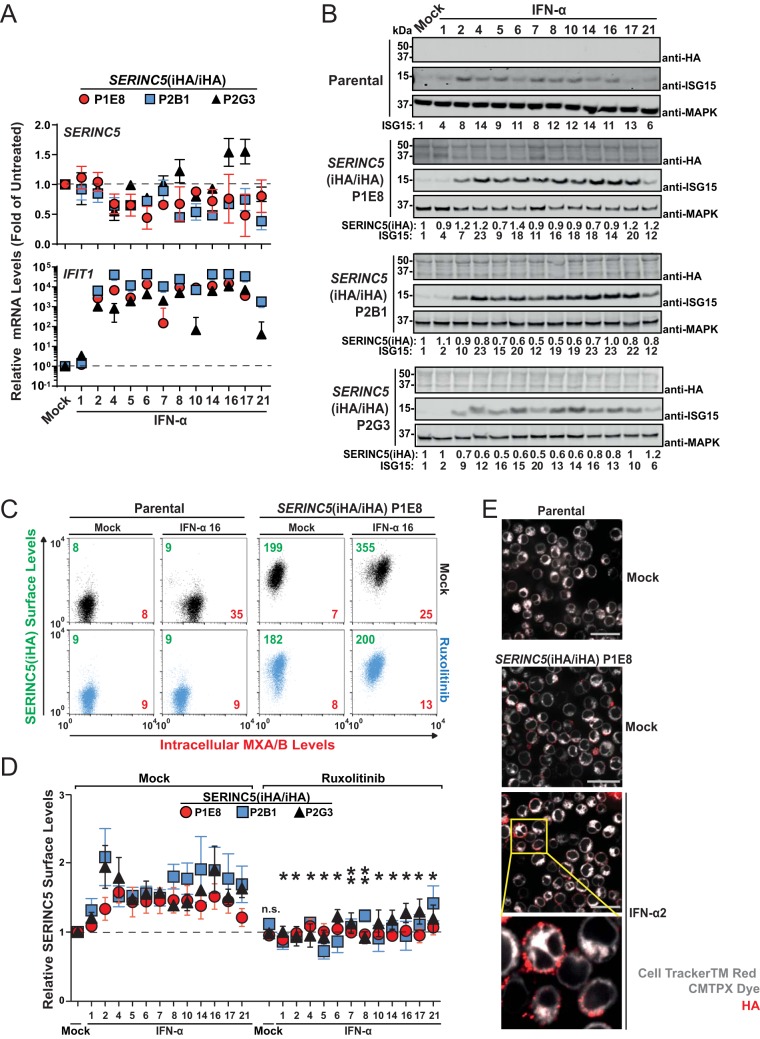
FIG 3.
Type I interferon modulates cell surface expression of endogenous SERINC5 protein in the absence of modulation of mRNA and protein quantities. (A) Q-RT-PCR analysis of SERINC5 and IFIT1 mRNA expression in SERINC5(iHA/iHA) Jurkat clones 48 h after treatment with the indicated IFN-α subtypes (10 ng/ml) or mock treatment. SERINC5 and IFIT1 mRNA expression levels in mock-treated cells were set to a value of 1. Error bars indicate SEM of data from three independent experiments. (B) Parental cells and the indicated SERINC5(iHA/iHA) clones were treated with the indicated IFN-α subtypes (10 ng/ml) or mock treated. Lysates were then subjected to anti-HA, anti-ISG15, and anti-MAPK immunoblotting. Numbers indicate fold changes in levels of the indicated proteins. For each cell line, one representative immunoblot from two to three independent experiments is shown. (C) Parental cells and the SERINC5(iHA/iHA) clone P1E8 were treated for 18 h with ruxolitinib (10 μM) or mock treated, followed by treatment with the indicated with IFN-α subtypes (10 ng/ml) or mock treatment for an additional 48 h. Cells were then immunostained with anti-HA for surface SERINC5(iHA), PFA fixed, permeabilized, and immunostained for intracellular MXA/B. Numbers indicate mean fluorescence intensities for SERINC5(iHA) (green) and for MXA/B (red). Shown are representative dot plots from one experiment out of six independent experiments. (D) Quantification of relative cell surface SERINC5(iHA) protein expression in the indicated SERINC5(iHA/iHA) clones. Error bars indicate SEM of data from two to six independent experiments, including the one shown in panel C. Statistical analysis refers to each individual interferon subtype in the absence and presence of ruxolitinib. (E) The SERINC5(iHA/iHA) clone P1E8 was treated for 48 h with IFN-α2a (250 U/ml), labeled with CellTracker red CMTPX dye, and immunostained with anti-HA. Cells were then PFA fixed and analyzed by confocal microscopy. Mock-treated parental cells are shown as a specificity control.

