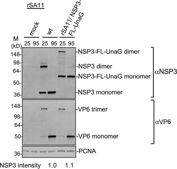
FIG 3.

Dimerization of NSP3-FL-UnaG. MA104 cells were mock infected or infected with rSA11/wt (wt) or rSA11/NSP3-FL-UnaG and incubated until 8 hpi, when cells were harvested. Cell lysates were mixed with sample buffer containing sodium dodecyl sulfate and β-mercaptoethanol, incubated for 10 min at either 25°C or 95°C, resolved by electrophoresis on a Novex 8 to 16% polyacrylamide gel, and blotted onto a nitrocellulose membrane. Blots were probed with guinea pig polyclonal anti-NSP3 or anti-VP6 antibodies or with a mouse anti-PCNA monoclonal antibody. Primary antibodies were detected using HRP-conjugated secondary antibodies. Sizes (kilodaltons) of protein markers (M) are indicated. NSP3 band intensities were determined by ImageJ analysis and were normalized to VP6 band intensities.
