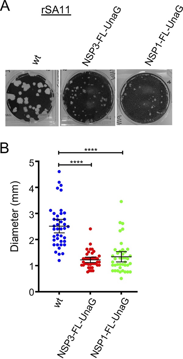
FIG 4.

Comparison of plaques formed by wild-type (wt) rSA11 and mutant rSA11 strains containing NSP3-FL-UnaG or NSP1-FL-UnaG sequences. (A) Plaques were generated on MA104 monolayers and detected at 6 dpi by crystal violet staining (39). (B) Sizes of 40 randomly selected plaques from six independent plaque assays were measured, and the means were determined. Mean values and 95% confidence intervals are plotted (black lines). Significance values were calculated using an unpaired Student's t test (GraphPad Prism v8). ****, P < 0.0001.
