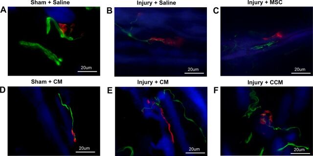
Fig. 4.
Examples of immunofluorescence of the EUS showing neuromuscular junctions (red), innervating nerves (green), and striated muscle (blue). Sham-injured animals demonstrated discrete organized motor endplates innervated by straight thick innervating axons (A and D). Three weeks after pudendal nerve crush and vaginal distension (Injury) with saline or CM treatment, innervating axons were thinner and motor endplates were less organized and diffuse (B and E). With MSC or CCM treatment, innervating axons took a more torturous course and had multiple collaterals (C and F).

