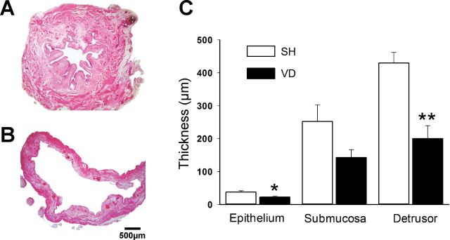
Fig. 4.
Photos of transverse sections of the bladder (hematoxylin and eosin stain) in SH (A) and VD (B) animals. Note that the thicknesses of the epithelium and detrusor layers were significantly decreased in VD animals (C). Values are means ± SE of data from 4 animals. *Significant difference vs. SH animals with P < 0.05; **significant difference vs. SH animals with P < 0.01.

