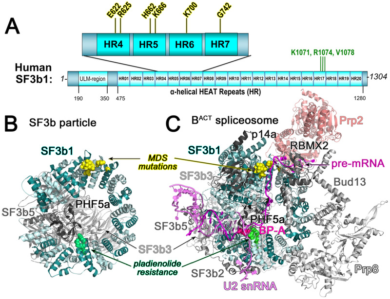
Figure 2.
(A) Domain organization of SF3b1. Recurrent cancer-associated mutations are expanded above (yellow highlight). Amino acids for which substitutions confer pladienolide-resistance are named (green font). HR, dihelical HEAT repeat; ULM, U2AF Ligand Motif. (B) Structure of the SF3b particle (PDB ID 5IFE). (C) Representative structure of the BACT spliceosome (PDB ID 5Z58 shown to illustrate intron position). SF3b1, cyan; U2 snRNA, violet; pre-mRNA, magenta. Mutational hotspots are yellow spheres; pladienolide-resistance sites are green spheres; branch point adenosine (BP-A) is shown as hot pink spheres.

