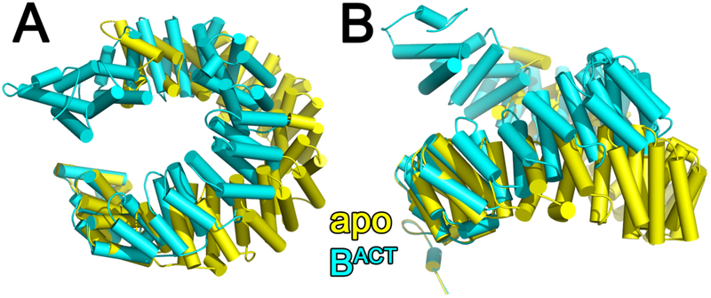
Figure 4.
(A) Comparison of human SF3b1 conformation in the isolated SF3b particle (yellow, PDB ID 5IFE) with the BACT spliceosome (cyan, PBD ID: 6FF4, a representative structure of the human BACT at relatively high resolution, Table 1) following alignment of the C-terminal repeat (residues 1244-1285). (B) View rotated 90° about the x-axis relative to (A). Results are similar following comparison of SF3b1 of the SF3b particle with other spliceosome structures. A movie portraying the conformational transition is given as supplementary information (Supplementary Movie S1).

